Mapping the Mind: Advances in Understanding Speech Production
(Posted on Monday, April 8, 2024)

This article was originally published on Forbes on 4/8/24.
This story is part of a series on the current progression in Regenerative Medicine. This piece discusses advances in neuroscience.
In 1999, I defined regenerative medicine as the collection of interventions that restore normal function to tissues and organs damaged by disease, injured by trauma, or worn by time. I include a full spectrum of chemical, gene, and protein-based medicines, cell-based therapies, and biomechanical interventions that achieve that goal.
Recent investigations of speech-related brain stimulation may point to how we plan what we say before we say it. A study by Kabakoff and colleagues in Brain Communications analyzes the regions of the brain that affect speech patterns and errors when electronically simulated. This knowledge could lead to more advanced speech therapies in the very near future.
The workings of the human mind have always been a subject of intrigue and fascination, and one of its most captivating functions is speech. Through speech, we convey our innermost thoughts, feelings, and concepts to others, and it serves as a medium of connection between ourselves and the world around us. However, the relationship between the mind and speech is complicated, and continues to be a subject of study and exploration.
For years, scientists have analyzed the complexities of speech production. While it is widely acknowledged that the cerebral cortex is a crucial component in speech, particularly in initiating the physical act of speaking, the planning phase of speech remained relatively unresearched. However, new data may lead to a better understanding of how we plan what we say.
A recent NYU Grossman School of Medicine study analyzed hundreds of brain-mapping recordings from 16 patients preparing for epilepsy surgery.
Before the operation, surgeons conducted a safety procedure to ensure that no vital brain regions were removed during surgery that could affect speaking ability. This procedure involved electrically stimulating specific parts of the brain while patients performed simple speaking tasks.
During these procedures, researchers observed the impact of stimulation on the speech production of a particular brain region, particularly the time it took for the stimulation to affect speech.
For instance, the surgeon may ask the individual to recite the Pledge of Allegiance, during which different areas of the brain would be stimulated, and the speech would be impacted, whether by slurring or total loss of speech.
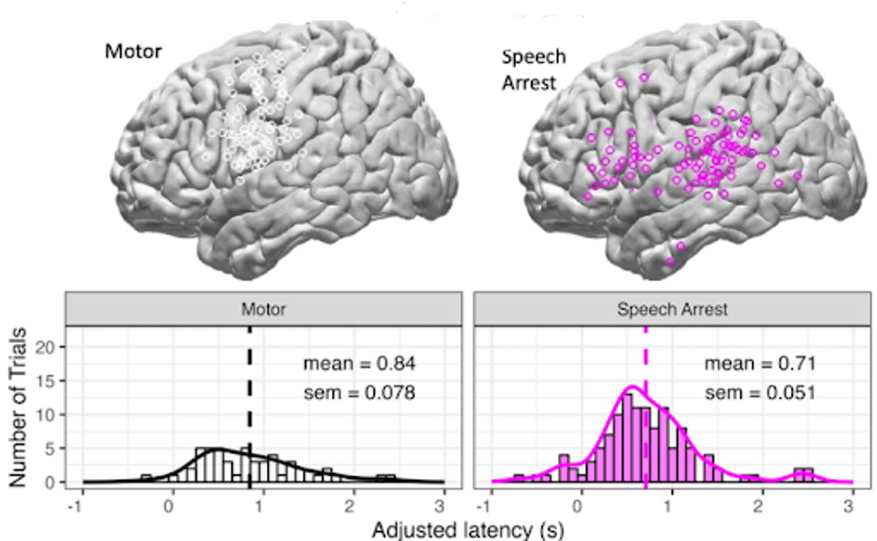
Stimulation would result in either motor-based arrest, which involves speech cessation due to vocal tract impairment, or speech arrest, which the researchers define as speech cessation that cannot be explained by motor interruption.

FIGURE 1: Cortical locations and distributions of latencies for all motor and speech arrest hits, controlling for trials with afterdischarges.
Stimulating the sensorimotor cortex caused most motor-based arrests (87.5%), whereas many different brain regions resulted in speech arrest.
Different brain regions also responded differently to stimulation. While some areas responded quickly, others took almost twice as long to impact the speech production process.

FIGURE 2: Map of the density of speech arrest hits across cortex in four time bins of adjusted latency.
Two adjacent regions of the cerebral cortex, namely the ventral sensorimotor cortex and the inferior frontal gyrus, have been found to exhibit longer latencies, between 0.5s and 1s, during speech planning. The ventral sensorimotor cortex is involved in the muscle movements necessary for speech production. In contrast, the inferior frontal gyrus involves various aspects of language processing, including grammar and syntax. Together, these two regions of the brain work in tandem to facilitate the complex process of speech production.
Conversely, smaller latencies of less than 0.5s were observed in other parts of the motor cortex and other gyrus, indicating that these regions play a more critical role in the physical mechanics of speaking.

FIGURE 3: Smoothed distributions of adjusted latencies, including speech arrest hits depicted with peaks in ascending order: supramarginal gyrus, STG, MTG, sensorimotor cortex, and IFG, and as a comparison, the motor hits in sensorimotor cortex.
These results are fascinating and may have profound implications in the near future. Brain mapping efforts work to plot the qualities and properties of brain function spatially.
Brain mapping is often used to diagnose neural impediments related to speech, vision, and movement. A more robust understanding of the human brain could improve techniques for diagnosing and treating neurological disorders and pave the way for more advanced neuroprosthetics to assist individuals with speech, vision, and movement impairments.
Understanding the neural mechanisms underlying speech production and language processing enables future scientists and engineers to develop more powerful AI-powered speech recognition and natural language processing technologies.
The future of neurological understanding and the cascade of effects that this understanding will entail, such as in neuroscience and neurotechnology, is bright. We live at the cusp of a new renaissance of regenerative medicine, bioelectronics, and medical devices—future advances that could aid the lives of thousands if not millions.

