A New Target to Boost Cancer Immunotherapy: LAG-3
(Posted on Tuesday, August 6, 2024)
Emerging research suggests that cancer therapies such as checkpoint inhibitors could be advanced by targeting a new protein. The protein, known as lymphocyte activation gene-3 or LAG-3, may prove particularly useful in improving patient responses to existing cancer treatments.
Killing Cancer with Checkpoint Inhibitors
Checkpoint inhibitors are an anticancer drug that indirectly attacks tumors. They depend on infusions of antibodies to block proteins called immune checkpoints. Specifically, they target one of three checkpoints: CTLA-4, PD-1 or PD-L1. The antibody barrier formed prevents checkpoint proteins from shutting down immune cells and allows the immune cell to do what it normally does: recognize and attack threats, including tumors.
These inhibitors can treat a wide range of advanced cancers—from lymphomas, lung cancers, skin cancers and more. They can even be combined with chemotherapy, radiation and other cancer treatments. However, some patients do not respond to the treatment at all, or may grow resistant to it over time.
Researchers believe that discovering new protein targets may improve patient responses to these therapies. The target may provide a new angle to attack tumors and therefore give a needed boost to existing checkpoint inhibitors.
LAG-3, An Experimental Checkpoint
Several new checkpoint proteins have been discovered since the development of current-day checkpoint inhibitors. A previous article discussed PD-L2, a potential companion to PD-1 and PD-L1 targeting checkpoint inhibitors. Another promising alternative is lymphocyte activation gene-3 or LAG-3.
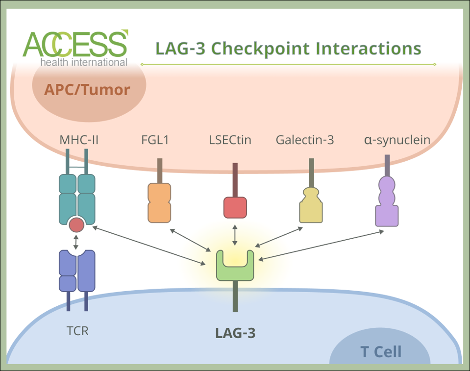
Lymphocyte activation gene-3 is a checkpoint protein found primarily on the surface of activated T cells. Several types of tumors can also express the protein, such as lung, breast and pancreas cancers. Despite its initial discovery in 1990, the exact mechanisms of action have yet to be uncovered. We do know that the receptor downregulates T cell function by binding to a selection of five other receptors. This starkly contrasts CTLA-4, PD-1 and PD-L2 checkpoints, which only interact with one or two partner proteins.
One known partner receptor is Class II Major Histocompatibility Complexes (MHC-II). Macrophages and other antigen-presenting cells rely on this protein to interact with and activate a subset of T cells. Yet, when LAG-3 binds to the complex, the opposite occurs; the checkpoint interrupts T cell activation and decreases necessary chemicals for the cell to survive and multiply. Notably, the checkpoint does not prevent the MHC II complex from binding to its normal partner receptor.
The checkpoint can also bind to fibrinogen-like protein 1 (FGL1), a protein typically expressed in the liver and pancreas at low concentrations. Evidence suggests that various tumors—lung, prostate, colorectal and other cancer cells—upregulate this protein to evade immune detection. Other partner proteins include LSECTin and Galectin-3, two receptors found on specific tumor cells, and at a lesser capacity Ɑ-synuclein, a receptor found on neurons in the central nervous system.

Figure 1: LAG-3 checkpoints interact with four known receptors on antigen-presenting cells (APCs) or tumor cells: class II Major Histocompatibility Complexes (MHC-II), Liver Sinusoidal Endothelial Cell Lectin (LSECtin), galectin-3, ɑ-synuclein, and fibrinogen-like protein 1 (FGL1).
ACCESS HEALTH INTERNATIONAL
Other Experimental Routes
LAG-3 targeting may also be possible via a different route: bispecific antibody therapy. This method relies on antibodies that can target two checkpoint proteins at once. Though these antibodies, in theory, accomplish the work of two inhibitors in one treatment, the hope is to exceed the tumor killing of dual-inhibitors—especially in cases of treatment resistance, when LAG-3 checkpoints may be upregulated.
For example, bispecific antibodies can bridge cells expressing PD-1 and LAG-3 while blocking these checkpoints. When this happens, the close cell-to-cell contact could encourage interactions that activate T cells. This bridging may also recruit other T cells to the site, potentially creating clusters of activated T cells.
Additionally, these antibodies may work more efficiently due to a phenomenon known as cross-arm avidity. In simple terms, when a bispecific antibody attaches to one checkpoint, it brings more antibodies into the area. This increases the chances of binding to the second checkpoint on nearby cells.
A bispecific antibody targeting PD-1 and LAG-3 has yielded encouraging response rates for patients with treatment-resistant solid tumors. Tumors decreased in 34% of patients when administered alone and 19% when given alongside an anti-HER2 cancer treatment.
Another alternative approach involves soluble fusion proteins. This method engineers an entirely new protein using components from LAG-3 checkpoints and immunoglobulin G antibodies. The fusion protein binds to MHC II complexes and activates antigen-presenting cells—in other words, they reverse the immunosuppressive signals sent by LAG-3 checkpoints. While still experimental, the protein has demonstrated clinical efficiency for late-stage breast cancer and advanced melanoma in combination therapies.
Looking Ahead
Cancer care continues to evolve, especially for checkpoint inhibitor research. Each new immune checkpoint discovery opens up possibilities for innovative treatment combinations. LAG-3 targeting inhibitors, a recent addition to the checkpoint inhibitor arsenal, have shown significant promise for patients with advanced melanoma since their approval in 2022. However, for now, the treatment must be administered alongside an anti-PD-1 targeting inhibitor. Ongoing research and clinical trials may uncover the underlying mechanisms of LAG-3 inhibitors, potentially broadening their therapeutic applications and enhancing outcomes for more patients in the future.
This article joins a growing series on mono cancer treatments, including novel immunotherapies such as CAR T therapy and checkpoint inhibitors. Find more at www.williamhaseltine.com
Read the original article on Forbes.

